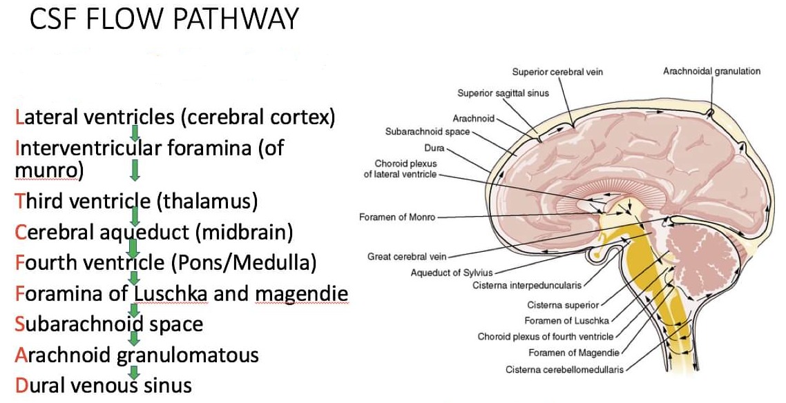
The four connected ventricles form chambers within the brain and are filled with CSF (Figure A and B).

- Lateral ventricles. These paired, C-shaped chambers are located deep within each cerebral hemisphere. Each lateral ventricle communicates via the interventricular foramen (of Monro) with the third ventricle.
- Third ventricle. The third ventricle is narrow and is located midline and inferior to the lateral ventricles. The third ventricle is positioned between the left and right diencephalon. The third ventricle communicates with the fourth ventricle via the cerebral aqueduct (of Sylvius).
- Fourth ventricle. The fourth ventricle is located superior to the pons and the medulla oblongata.

A. Three-dimensional lateral view of the ventricles of the brain. B. Coronal section of the brain showing the ventricles. C. Formation, location, and circulation of cerebrospinal fluid (CSF).
Cerebrospinal Fluid
CSF is produced by the choroid plexuses, located within each of the ventricles, and flows from the lateral and third ventricles to the fourth ventricle via the cerebral aqueduct. From the fourth ventricle, CSF enters an enlarged part of the subarachnoid space (cisterna magna) via the central median aperture (of Magendie) and the lateral apertures (of Luschka). The CSF circulates around the spinal cord and brain in the subarachnoid space to empty into the superior sagittal sinus via the arachnoid granulations. Arachnoid granulations are projections of the arachnoid mater along the superior sagittal sinus. The brain and spinal cord are very delicate and, as such, are susceptible to damage. Therefore, the brain and spinal cord are well protected through the bony encasement (skull and vertebral column) as well as by a second protective layer of connective tissue coverings (the meninges). The third protective mechanism is CSF, which supports the brain and spinal cord. Despite all of these protections, trauma to the brain and spinal cord can still occur and can result in devastating injuries and deficits.
The lumen of the cerebral aqueduct or the fourth ventricular apertures may become obstructed. When either of these conditions occurs, CSF continues to be secreted, producing excessive pressure within the ventricles. In children, this results in hydrocephalus, a condition in which the head enlarges because the skull bones have not yet fused. In adults, however, hydrocephalus is a different challenge because the skull is rigid. Therefore, the accumulating CSF compresses the brain tissue. In most cases, hydrocephalus is treated by inserting a shunt into the ventricles to drain the excess CSF into either a jugular vein or the peritoneal cavity.